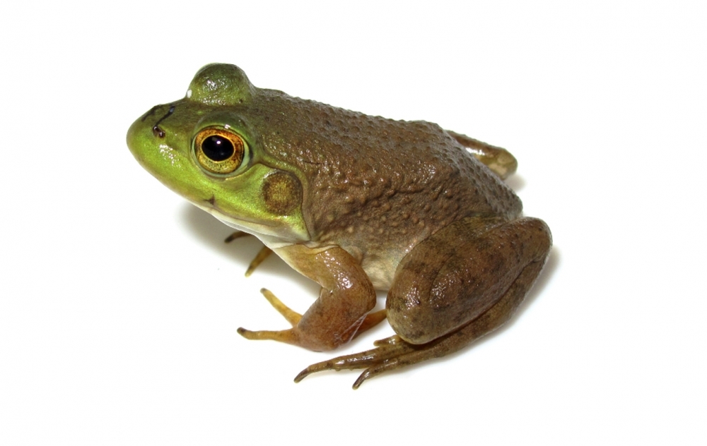


Ranavirus is linked to amphibian decline or extinction in other parts of the world, but in Brazil, it has been reported only in captive animals (bullfrog; photo: Felipe Toledo)
Published on 05/12/2021
By Maria Fernanda Ziegler | Agência FAPESP – Researchers have found bullfrog tadpoles with clear signs of infection by ranavirus in Brazil. The specimens were collected from two ponds in the city of Passo Fundo, South of the country (state of Rio Grande do Sul), in November 2017. Ranavirus causes skin ulcerations, edema and internal hemorrhage. It does not affect humans but can be lethal to amphibians and fish.
This is the first infection of wild amphibians detected in Brazil. “The discovery causes concern, as it’s the first time ranavirus has been found in nature here. Epidemics were reported in 2006 and 2009, but they occurred at frog farms, not in the wild. The virus has been detected in nature elsewhere in the world and is associated with the decline in populations of amphibians, Earth’s most endangered group of vertebrates,” said Joice Ruggeri, a researcher at the University of Campinas’s Biology Institute (IB-UNICAMP) and first author of the article published in the Journal of Wildlife Diseases.
The discovery resulted from Ruggeri’s postdoctoral research, supported by FAPESP. The aim of the project was to investigate the dynamics of ranavirus in different species of anurans (frogs and toads), its interaction with other pathogens, and possible imbalances or threats to populations of these animals in the Atlantic Rainforest biome.
Ruggeri collected specimens of wild anurans in areas from Rio de Janeiro state in Southeast Brazil to Rio Grande do Sul in the South, including ponds in Passo Fundo.
“We found many dead tadpoles and fish in these ponds. It was a scene of destruction. We’re now analyzing all the data collected, which should provide interesting answers regarding the relations among the different pathogens that are threatening anuran populations in the Atlantic Rainforest,” Ruggeri told Agência FAPESP.
The discovery also raises questions about the relations between invasive and native species. According to the article, the researchers found dead and dying adults and tadpoles of both native species and the American bullfrog (Lithobates catesbeianus), an invasive species. All had been infected by the virus.
L. catesbeianus can be infected with ranavirus without contracting any disease, thereby acting as a vector for its dissemination. This reinforces the hypothesis, as yet unconfirmed, that native species are being infected by the invasive species.
The weight of invasive species
L. catesbeianus is native to North America and has been introduced to over 40 countries on four continents. It is one of the main species farmed for human consumption. Brazil is the second-ranking producer. Most farms are located in areas of Atlantic Rainforest between Rio de Janeiro and Rio Grande do Sul, rationalizing the researchers’ choice of these areas for data collection.
“Frog breeding as a business has ups and downs. Several frog farms have been abandoned since 1990, and many animals escaped into the wild as a result,” said Luís Felipe de Toledo, a professor at IB-UNICAMP and one of the authors of the article.
The researchers believe the virus may have spread through the Atlantic Rainforest biome from frog farms, but there are other hypotheses. “We know many of these frogs escaped from captivity and may have taken the virus into the natural environment, but we’re not yet sure whether they have any link to the virus detected in species native to Brazil,” Ruggeri said.
Dual threat
The discoveries do not stop there. Two of the bullfrogs studied had been coinfected by ranavirus and by the fungus Batrachochytrium dendrobatidis, also known as Bd or amphibian chytrid fungus, which causes chytridiomycosis and is responsible for the greatest biodiversity loss due to a single pathogen ever recorded.
In an article published in Science in March 2019, researchers from 16 countries, Toledo among them, reported that the fungus has caused a decline in the populations of at least 501 species of amphibians in the past 50 years.
“It’s one more threat to these animals. The fungus has already been linked to amphibian extinctions here in Brazil. Now that we’ve found cases of infection by ranavirus, we wonder if it hasn’t also caused declines or extinctions,” Toledo said.
Both fungi and viruses are transmitted via exposure to infected water or direct contact between frogs or tadpoles, making dispersal of these microorganisms highly effective. While the fungus disrupts the balance of fluids and electrolytes, ultimately leading to cardiac arrest, the virus can cause cell death in multiple organs.
“When an amphibian is infected by both pathogens, it becomes even more vulnerable. However, our understanding of the consequences of this coinfection is still insufficient,” Ruggeri said.
Having analyzed the data collected, the researchers will now investigate the ranavirus strains found in South Brazil. “The virus’s lineage could be Brazilian. We also plan to determine whether the viruses identified in the wild and at frog farms are from the same lineage. The discovery points to several lines of research,” Toledo said.
High mortality
Low levels of ranavirus were detected in native tadpoles living in one of the ponds, which contained no bullfrog tadpoles.
More than 20 dead bullfrog tadpoles were found in the other pond, and no native anuran species were found. All the animals found had severe skin lesions and were infected by ranavirus. Only two tadpoles were found alive, and these had low levels of infection.
The fungus was detected in seven out of 19 dead tadpoles, including samples of native and invasive species collected from both ponds. However, the researchers ruled out chytridiomycosis as the cause of death because the fungal load was low.
Although bullfrogs are generally tolerant of ranavirus, high viral loads were found in their blood, and they displayed clear clinical signs of disease.
The article “First case of wild amphibians infected with ranavirus in Brazil” (doi: 10.1038/s41467-019-08909-4) by Joice Ruggeri, Luisa P. Ribeiro, Mariana R. Pontes, Carlos Toffolo, Marcelo Candido, Mateus M. Carriero, Noeli Zanella, Ricardo L. M. Sousa and Luís Felipe Toledo can be read at www.jwildlifedis.org/doi/pdf/10.7589/2018-09-224.
Source: https://agencia.fapesp.br/30808