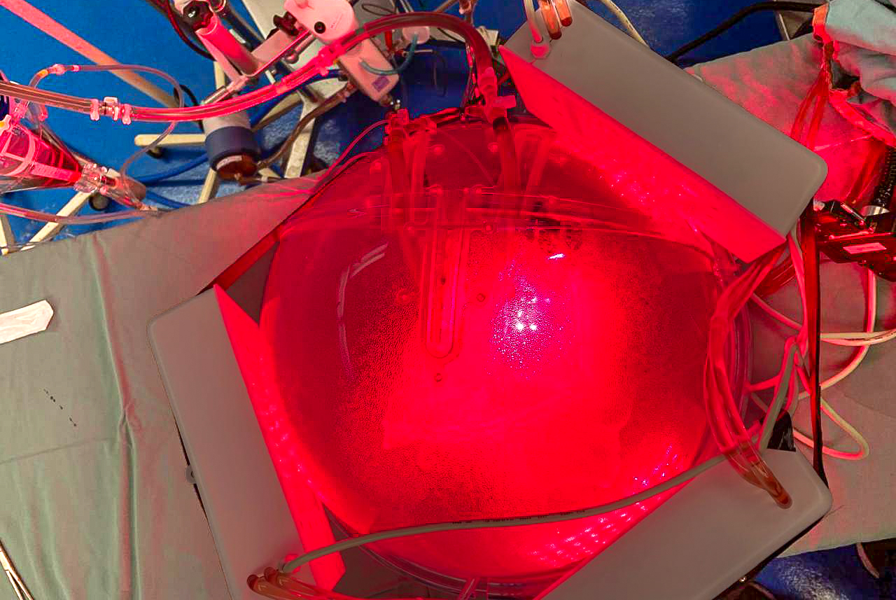
Light-based therapy applied to a lung intended for transplantation. The same type of light source can be used to treat pneumonia (image: Cristina Kurachi)
Published on 10/19/2026
By José Tadeu Arantes | Agência FAPESP – Researchers at the University of São Paulo (USP) in Brazil and their international collaborators have created a computational model that can generate a “digital twin” of the human thorax. The goal is to personalize the treatment of respiratory diseases using a special type of light. Described in a paper published in PNAS, the journal of the U.S. National Academy of Sciences, the new technology uses CT scans to predict the path that light radiation will take inside the body. This makes it possible to calculate the exact dose needed to combat infections and inflammation directly in the lungs by simply illuminating the person from the outside.
Light-based therapies have been used to treat inflammation or infections in various organs. The two main approaches are photodynamic therapy, which uses light to activate a photosensitive substance that generates reactive species capable of destroying microorganisms in the presence of oxygen, and photobiomodulation, which uses low-intensity light to modulate cellular processes and inflammatory responses. However, in the case of the lungs, the use of these techniques presents a significant challenge. To reach the organ noninvasively, the externally applied light must pass through the skin, fat, muscles, bones, and other chest tissues, undergoing absorption and scattering along the way.
Thus, the same amount of light applied to two people’s chests can produce very different effects in their lungs. To address this issue, the authors combined computed tomography images, three-dimensional anatomical reconstructions, and computational simulations accelerated by high-performance computing. This process created a “digital twin” of each patient’s chest. This information makes it possible to estimate how much light will reach different regions of the lungs and which configuration would be most appropriate for each anatomy and pathological condition.
“Since our strategy uses external illumination, we need to know how much light reaches the lung and if the dose is sufficient to activate the drug in photodynamic therapy or induce the biological response in photobiomodulation,” says Cristina Kurachi, a professor at the São Carlos Institute of Physics at the University of São Paulo (IFSC-USP) who coordinated the study alongside Vanderlei Bagnato, also from IFSC-USP. Researchers from the Hospital das Clínicas of the USP Medical School (HC-FM-USP), Texas A&M University in the United States, and Victoria University of Wellington in New Zealand also participated.
As Bagnato points out, the lung is a particularly difficult target for light-based therapies. One alternative would be to deliver light to the organ via bronchoscopy, which uses a thin tube with a camera (a bronchoscope) that is inserted through the nose or mouth. However, the procedure is invasive and may worsen the patient’s condition.
“External illumination is much simpler. However, in addition to the absorption and scattering that light radiation undergoes on its way to the organ, the lung itself isn’t a homogeneous medium. Air, lung tissue, blood vessels, and regions altered by inflammation or collapse have different optical properties. Focal pneumonia [concentrated in a specific region], diffuse pneumonia [in various regions of both lungs], and COVID-19, for example, alter the distribution and density of lung tissues in distinct ways,” Bagnato explains.
This complexity makes the idea of applying the same light intensity to all patients, for the same duration and in the same position, inappropriate in principle.
“Our solution was to use patient images, incorporating the anatomy of the tissues and their varying thicknesses, to create a ‘digital twin.’ In it, we included the optical properties of the tissues – that is, how much light is absorbed and how much is scattered – to try to predict the amount that actually reaches the lung and thus result in individualized dosimetry and a more effective therapeutic response,” Kurachi explains.
The first step involves converting the images obtained by computed tomography into a three-dimensional model of the patient. The tomography volumes are segmented, meaning the computer identifies and separates the various anatomical structures. In the study, 12 types of tissue and structures were represented: skin, subcutaneous fat, muscles, bones, heart, liver, stomach, kidneys, and in the lungs, normally ventilated tissue, collapsed regions, infiltrates, and blood vessels.
This anatomy is divided into small volumetric elements called voxels, which are the three-dimensional equivalents of pixels in a digital image. In the model used, each voxel measured 0.1 centimeter on each side. The result is a detailed computational representation of the thoracic cavity. Then, each tissue is assigned optical parameters, specifically the coefficients of light absorption and scattering at different wavelengths.
“With all this data incorporated into the model, Monte Carlo simulation comes into play. Rather than attempting to find a single analytical solution for light propagation in extremely heterogeneous anatomy, the simulation calculates the fate of millions of photons as they travel, scatter, or are absorbed by different tissues,” Bagnato explains.
Monte Carlo simulation is a computational method that uses repeated random sampling to obtain numerical solutions to complex problems involving many variables and probabilistic behaviors.
“Based on the collective behavior of the photons, the system constructs three-dimensional maps of the distribution of light and the energy absorbed within the body. In the study, the platform was fed CT scans of people with various pulmonary conditions. The group included four individuals from HC-FM-USP: one control subject without lung disease and three patients with predominantly focal bacterial pneumonia, diffuse bacterial pneumonia, and viral pneumonia caused by COVID-19, respectively. Two other volunteers participated in optical measurements used for the experimental validation of the model. The CT images were used to construct the ‘digital twin,’ and light distribution measurements were obtained from the volunteers, which allowed us to assess the model’s performance,” Kurachi explains.
The reconstructions clearly showed the extent to which disease alters the organ. In the healthy patient used as a reference, approximately 91% of the organ was normal tissue capable of filling with air to facilitate respiration. However, in the patient with focal pneumonia, the healthy area decreased to 58.9%. In the case of diffuse pneumonia, the healthy area was even smaller, at just 54.1%. The patient with COVID-19 showed a significant loss of lung function, retaining only about 69.9%. This lost functional space was eventually taken up by inflammation, collapsed areas that can no longer facilitate gas exchange, and an increase in blood vessels. These major physical changes make the organ denser and alter its interaction with light.
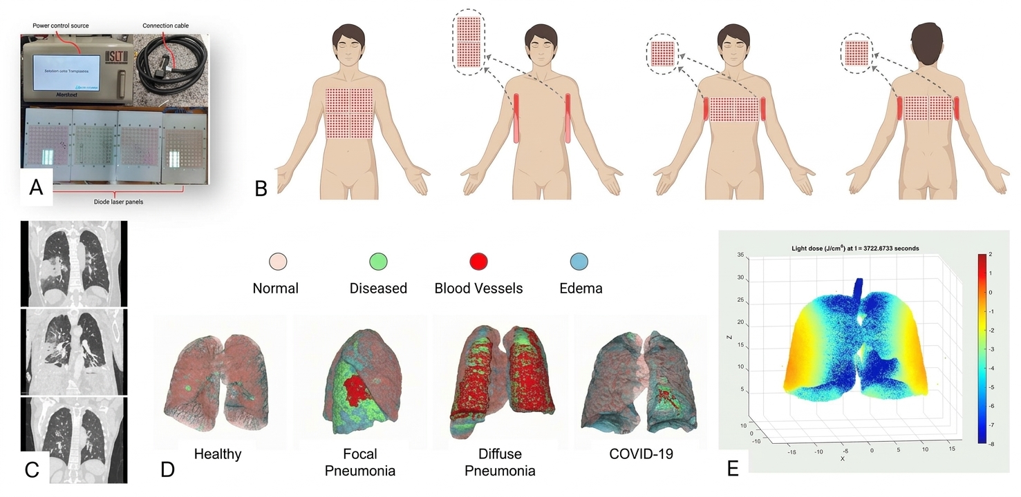
A) Lighting device for pulmonary phototherapy. B) Schematic diagram of lighting arrangement options. C) Computed tomography images used to tailor the lighting plan for each patient, taking into account chest anatomy and lung disease. D) Three-dimensional mapping of the patient’s lung conditions. E) Final result of the computational model showing the “digital twin” of the lungs with the light dose delivered to each region as a function of illumination time (figure: Cristina Kurachi)
The position of the light source makes a difference
Initially, different wavelengths and four configurations for positioning the light sources around the chest were evaluated. One result stood out: light at 808 nanometers, which is in the near-infrared spectrum and is therefore invisible to the human eye, showed the highest energy deposition in the lungs across all configurations tested.
The position of the light sources was also crucial. A configuration that illuminated the lateral and dorsal regions of the chest provided the most uniform distribution and highest energy deposition in the lungs. In contrast, frontal illumination alone performed poorly, mainly due to attenuation caused by the ribs and, in the anterior region of the chest, by breast adipose tissue.
“If the patient has early-stage pneumonia localized to one region of the lung, why illuminate the entire lung or both lungs? If the pneumonia is severe and diffuse, the situation is different. Based on the characteristics of the light source, the patient, and the disease itself, we determine which panels need to be turned on and how the illumination should be configured. We no longer simply illuminate without knowing the exact outcome. Instead, we tailor the planning to each individual case,” Kurachi emphasizes.
Of the simulations evaluated, the multi-angular configuration tested at 808 nanometers produced the most uniform and efficient distribution. This configuration involves positioning light sources around the patient to illuminate the chest from multiple directions simultaneously, combining the front, back, and sides of the body.
The differences observed in the simulations were not solely due to each patient’s anatomy. The disease itself alters the structure of the lungs, consequently altering the way tissues interact with light and the amount of light energy they absorb. The portion of light energy absorbed by the tissue is referred to by the researchers as “energy deposition.”
In the simulations, regions of collapsed lungs – where the alveoli lose air and the tissue becomes denser – exhibited the highest energy deposition proportionally, ranging from 3% to 4.5%. In normally ventilated lung parenchyma, the region composed of millions of alveoli where gas exchange occurs, this value was around 1% to 2%. Regions infiltrated by inflammation showed intermediate values. This occurs because changes in tissue density and composition alter light absorption and scattering properties. “From the perspective of treatment using photonic techniques, this behavior is positive since we want the greatest deposition and absorption of energy precisely in the regions where the disease is established while preserving normal lung tissue,” Kurachi notes.
These differences produced characteristic patterns for each clinical condition. In focal pneumonia, the greatest energy deposition was concentrated in the affected areas, forming well-defined zones that were distinct from healthy tissue. In diffuse pneumonia, energy was distributed more homogeneously across extensive regions of the lung. In cases of COVID-19, a bilateral and peripheral pattern emerged, corresponding to the distribution of opacities observed on CT scans.
“This study aims precisely to prevent patient radiation therapy planning from being done ‘in the dark,’ that is, placing radiation in a certain location without knowing how much will actually reach the target. The goal is to tailor the treatment plan based on the patient’s characteristics and the specific disease,” Kurachi summarizes.
How much light actually reaches the lungs?
Calculations showed that only a small fraction of the light applied to the skin reaches the lungs. Using 808-nanometer infrared light with an irradiance of 100 milliwatts per square centimeter (mW/cm²) on the skin yielded the best results. The average irradiance in a healthy lung was only 0.0234 mW/cm². In diseased lungs, the irradiance was even lower: 0.0166 mW/cm² in focal pneumonia, 0.0120 mW/cm² in diffuse pneumonia, and 0.0113 mW/cm² in patients with COVID-19. These conditions require long illumination times in a clinical setting, but intervals ranging from 40 to 80 minutes are sufficient to deliver the necessary energy for photobiomodulation and photodynamic therapy.
This is because much of the light is absorbed or scattered by the tissues it encounters along the way. The amount that reaches the lungs varies from person to person, as well as between different regions of the organ itself. This variation depends on factors such as the thickness of the chest wall, the geometry of the lungs, and changes caused by the disease.
“This is precisely where the digital twin can make a difference. We can use everything we’re able to observe in the patient to plan what we can’t observe directly. The goal is to get as close as possible to the required light distribution and make the treatment more precise,” Bagnato emphasizes.
The researchers have a precise plan for a future application. First, a high-resolution CT scan or MRI would produce a three-dimensional image of the patient’s anatomy. Healthy and diseased regions would then be identified. Next, the model would be assigned the optical properties corresponding to the different tissues.
Then, computer simulations would calculate how light propagates inside the chest in question. Before performing the actual procedure, the physician could virtually test different wavelengths, intensities, and source positions to choose the most appropriate configuration. Thus, the “digital twin” would function as a computational testing ground for the treatment.
However, it should be noted that the model is not yet ready for clinical use. The optical properties attributed to the different tissues were primarily obtained from the literature and do not account for all individual variations. Additionally, while the simulation results align with experimental measurements, further validation of the amount of light reaching the interior of the lungs is necessary.
Progress toward clinical application has already begun, though. “We’re conducting an in vivo study on sheep in the United States. Sheep develop pneumonia similar to that seen in humans, including cases caused by the same bacterium. This is a preparatory step toward moving on to human subjects,” Bagnato explains.
The next steps include further experimental and clinical validations, as well as real-time measurement of the light dose in the future. The goal is to develop personalized treatments that determine how much light to apply to the skin and how much should reach the intended region of the lung.
However, for Bagnato, technological sophistication must go hand in hand with another concern. “We’re scientists committed to the relevance of our research. We want to solve problems, but we also operate with social responsibility. We don’t want to create technology that’s too expensive, can’t be integrated into the Unified Health System [SUS], or used widely. A good treatment must be accessible to the population. The economic reality must be taken into account in what we're developing,” he says.
The study was funded by FAPESP through the Optics and Photonics Research Center (CEPOF) and the Thematic Project “Advanced Photonic Technologies for the Life Sciences”, both of which are coordinated by Bagnato.
The article “Anatomy-resolved digital twin framework for personalized light dosimetry in lung therapies” can be read at pnas.org/doi/10.1073/pnas.2612370123.
Source: https://agencia.fapesp.br/59424