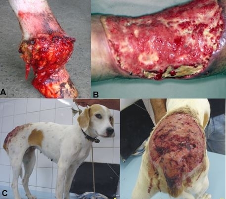
Ulcerations caused by pythiosis: (A) in the hind leg of a horse; (B) in a human ankle; C) in a dog (photos: researchers’ archive)
Published on 03/21/2022
By André Julião | Agência FAPESP – In the classes she teaches at São Paulo State University (UNESP) in Botucatu, Brazil, Professor Sandra de Moraes Gimenes Bosco likes to say that pythiosis is a tropical disease so neglected that it is even omitted from the list of neglected tropical diseases (NTDs) published by the World Health Organization (WHO), which comprises 20 NTDs that affect mainly the poor and for which effective treatment is not available. Pythiosis causes severe skin lesions and occurs in both humans and animals such as horses. Bosco’s research group has now increased the visibility of the disease by identifying seven potential antigens that can facilitate diagnosis and treatment.
The study was supported by FAPESP. Its findings are reported in an article published in the Journal of Fungi.
“We identified seven antigens commonly found in the blood serum of horses and humans who have had pythiosis. They’re proteins with high antigenicity, meaning they’re very likely to be recognized by the immune system. In addition, in computer simulations these molecules were detected by B lymphocytes, immune system cells responsible for producing antibodies, so they’re promising markers for diagnosis of the disease,” said Jéssica Luana Chechi, first author of the article. The study was conducted during her doctorate at the Botucatu Institute of Biosciences (IBB-UNESP), with Bosco acting as her thesis advisor.
“Pythiosis diagnosis, treatment and prognosis are all complex,” said Bosco, last author of the article. “It can be misdiagnosed as an infection by a class of fungi called zygomycetes, because the organisms are morphologically similar. The organism that causes pythiosis doesn’t respond to existing antifungal medications, so it can only be treated by surgical excision of all the infected tissue. Depending on the region affected, there isn’t much margin for surgery and the outcome can be amputation for humans or euthanasia for horses.”
The study was part of a project supported by FAPESP and led by Bosco. Chechi conducted part of it while she was on a research internship at Mahidol University in Thailand.
False fungus
Pythiosis is caused by Pythium insidiosum, an oomycete or false fungus. It looks like a fungus under the microscope, and there are other similarities in reproduction and life cycle, but it is more properly defined as a close relative to brown algae and microalgae. Hence the reason why pythiosis is not cured by the antifungals available on the market. In its systemic form, the disease can cause arterial occlusion (partial or total blockage) and death.
P. insidiosum lives in still water. Its zoospores (reproductive cells) penetrate the tissue of aquatic plants or existing wounds in mammals, where its life cycle continues. In Brazil, it is fairly common in horses, especially in the Pantanal (Center-West region), although it is also found in other regions. In 2019, for example, five horses were infected in a nine-day outbreak on rural properties in the area of Botucatu, near the Tietê River.
Curiously, the first case of equine pythiosis reported in Thailand occurred only in 2018. In that Southeast Asian country, pythiosis is considered an occupational disease because it mainly affects rice growers, causing leg wounds that can require amputation. The only case in humans reported in Brazil to date occurred in 2002, although the researchers suspect others have occurred but were not reported owing to ignorance of the disease among health workers.
Bosco herself first heard about pythiosis only in 2002 when she was finishing her PhD research. One day, a sample of a surgical piece removed from a man with a severe leg wound came to the laboratory where she conducted her research. He was a patient at the general and teaching hospital run by Botucatu Medical School (FMB-UNESP). Various treatments had been tried. Amputation had been considered, but the infection had been controlled after major surgery in the region.
“We thought a fungus was in the sample, but when we sequenced a specific DNA region and compared it with a database, the result pointed to P. insidiosum,” Bosco recalled. “That’s when my interest in it began.” Her first and master’s degrees were in veterinary medicine, and over the years she has diagnosed the disease in horses and dogs.
Besides ignorance of the disease, another obstacle to diagnosis is the lack of antigens (proteins that serve as biomarkers of infection). Only after a series of costly and time-consuming laboratory tests can the infectious agent be identified with precision.
Antigens
To find antigens in their latest study, the researchers first isolated P. insidiosum from the wounds of infected horses. Next, the oomycete was exposed to blood serum from 22 horses in Brazil and ten humans in Thailand. Both groups were diagnosed with the disease.
In trials of this kind, the antibodies created by prior infection are expected to bind to proteins in the infectious agent. Sera from healthy horses and humans were used as control, in order to ensure that the proteins found were specific to infection by the false fungus.
Several antigens responded to the isolate, but seven drew the researchers’ attention because they were recognized in both horses and humans, among other reasons. The proteins identified include several that play a key role in various biological processes involving the cellular stress associated with temperature changes. Others are essential to the development of P. insidiosum owing to the role they play in cell wall formation. Two of the antigens interact with macrophages (defense cells).
The researchers in Botucatu now plan to purify some of these proteins in order to test their potential as diagnostic markers or even for the development of a pythiosis vaccine. One such vaccine is already being developed at the Federal University of Santa Maria (UFSM) in partnership with EMBRAPA Pantanal, a unit of the Brazilian Agricultural Research Corporation (EMBRAPA). It is called Pitium-Vac, but it is designed for therapeutic use and not to prevent the disease. It is obtained from a macerate of the oomycete, and hence with total antigens rather than specific antigens like those found in the study.
“In an ideal world, we’d have an immunochromatographic test that could diagnose the disease with a drop of blood. A lot more research will be needed to get there, but we’ve taken the first step,” Chechi said.
The article “Prospecting biomarkers for diagnostic and therapeutic approaches in pythiosis” is at: www.mdpi.com/2309-608X/7/6/423/htm.
Source: https://agencia.fapesp.br/38199
