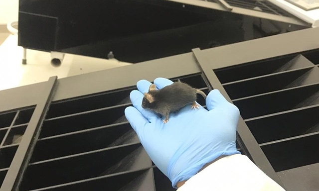
Studies show that in addition to skeletal muscle, the heart, liver and central nervous system are affected. The systemic action of proinflammatory cytokines explains only part of the phenomenon (photo: researcher's archive)
Published on 05/12/2021
By Karina Toledo | Agência FAPESP – Elite athletes frequently end up underperforming when they train heavily without sufficient rest and recovery, owing to what is known as overtraining syndrome. Symptoms may include depression, weight loss, insomnia, irritability, diminished immunity and loss of appetite.
Until now, the most widely accepted explanation for the phenomenon has been that injuries to skeletal muscle tissue due to overtraining lead to an increase in the release of cytokines (immunomodulatory proteins secreted by cells) and other proinflammatory substances into the bloodstream, triggering systemic effects. The theory was put forward two decades ago and is known as the cytokine hypothesis.
A series of studies conducted more recently at the University of São Paulo (USP) in Ribeirão Preto, Brazil show that the effects of overtraining on the organism go far beyond performance decline and can cause lasting damage to the heart, liver and central nervous system, as well as skeletal muscles. Experiments with mice have also refuted the hypothesis that proinflammatory cytokines are the only factor responsible for the drop in performance. The mice continued to underperform after blood levels of these substances returned to normal.
These studies have been conducted over the past ten years with FAPESP’s support. The principal investigator is Adelino Sanchez Ramos da Silva, a professor at the University of São Paulo’s Ribeirão Preto School of Physical Education and Sports (EEFERP-USP). The main findings, as well as data from studies by other research groups, are described in a review article published in the journal Cytokine.
“This information should serve as a warning against overtraining. Elite athletes often have no choice, owing to pressure from coaches, sponsors and competitions, but the need for a minimum of recovery time should at least be respected,” Silva said in an interview given to Agência FAPESP.
Overtraining protocols involving downhill, uphill and level running were tested in mice by Silva and his team with the aim of understanding the action of proinflammatory cytokines induced in different tissues by excessive physical exercise. In all cases, the training period was eight weeks, with the first four being devoted to adaptation.
“In the seventh week of the protocol, the mice were made to run on a treadmill at an intensity equivalent to 75% of their previously measured maximum capacity. Training sessions lasting 75 minutes took place once a day from Monday through Friday. In the eighth week, the mice ran twice a day, with a four-hour interval between sessions,” Silva said.
All three overtraining protocols produced an increase in blood serum levels of three proinflammatory cytokines: interleukin-1-beta (IL-1β), interleukin-6 (IL-6) and tumor necrosis factor-alpha (TNF-α). The increase in proinflammatory molecules was also observed in skeletal muscle tissue, with different effects for each protocol.
The deleterious effects of downhill overtraining were the most severe, including skeletal muscle atrophy and endoplasmic reticulum stress. The endoplasmic reticulum is a subcellular organelle that ensures that proteins fold into their correct shapes, among other functions. “Put simply, this means cells in the region ended up containing misshapen proteins, which could impair cellular functioning,” Silva explained.
After two weeks of rest, the serum and skeletal muscle proinflammatory cytokines had returned to baseline levels, and intramuscular anti-inflammatory cytokines had increased. “However, the animals’ running performance did not return to normal. It was still diminished, suggesting that other mechanisms were involved in the process,” Silva said.
The downhill overtraining protocol caused loss of weight and appetite, as well as inflammation of the hypothalamus, a brain region that regulates several metabolic processes.
“All three protocols induced endoplasmic reticulum stress in the hypothalamus, but we didn’t observe cell death,” Silva said.
Central nervous system inflammation was reversed after the two-week recovery period. Body weight and appetite also returned to normal.
Insulin resistance and cardiac hypertrophy
All three overtraining protocols led to impairment of the insulin signaling pathway in skeletal muscle, so that the muscle cells were less able to draw glucose from the bloodstream. Nevertheless, glucose tolerance testing did not point to negative alterations, showing that blood sugar was properly metabolized by the organism.
“We suspected this balance was maintained because some other tissue could be compensating for the skeletal muscle insulin signaling pathway impairment. Our analysis did in fact show an improvement in the liver insulin signaling pathway and an increase in hepatic glycogen accumulation in the animals subjected to the downhill and uphill overtraining protocols. As a negative adaptation, however, we observed an accumulation of fat and signs of liver tissue inflammation,” Silva said.
The results also suggested that the heart acts to compensate for the impairment of glucose capture by skeletal muscle cells, as glycogen accumulation in cardiac tissue was observed in response to all three overtraining protocols. Surprisingly, the researchers also found signs of fibrosis in the left ventricle. Only the mice subjected to the downhill running protocol showed molecular signs of pathological cardiac hypertrophy.
“In the studies performed to date, we did not measure functional cardiac parameters. We’re now reproducing the experiments and will perform an echocardiogram at the end of the training period to determine whether the left ventricle is functionally impaired,” Silva said.
The group also plans to investigate whether cardiac and hepatic molecular alterations are reversed after the two-week rest. “I imagine this is the case, but we haven’t looked into it yet,” Silva said.
Generally, the downhill protocol caused more damage. According to Silva, this was due to the predominance of eccentric contractions during downhill running. In an eccentric contraction, a muscle lengthens while undergoing tension because the external force exceeds that produced by the muscle.
Future studies
The studies conducted on USP’s Ribeirão Preto campus were supported by FAPESP via three regular research grants awarded to Silva, in 2010, 2014 and 2018.
Other grants awarded by FAPESP in connection with the studies went to Ana Paula Pinto for research in connection with her master’s degree and PhD; to Alisson Luiz da Rocha for scientific initiation, master’s research and PhD research; and to Bruno Cesar Pereira for master’s research and scientific initiation.
Although the group’s findings do not support the hypothesis that proinflammatory cytokines are the only factor responsible for the decline in physical performance, interleukin-6 (IL-6) was involved in most of the effects observed in the organs studied after all three overtraining protocols.
In recent experiments, the researchers observed that in IL-6 knockout mice (genetically modified so as not to express this cytokine), skeletal muscle endoplasmic reticulum stress after a single intense training session was milder than that in wild-type (non-GM) mice.
“Future studies will have to investigate better the role of this cytokine in the metabolism as part of the effects of overtraining,” Silva said.
The article “The pro-inflammatory effects of chronic excessive exercise” by Alisson L. da Rocha, Ana P. Pinto, Eike B. Kohama, José R. Pauli, Leandro P. de Moura, Dennys E. Cintra, Eduardo R. Ropelle and Adelino S.R. da Silva can be retrieved from: www.sciencedirect.com/science/article/abs/pii/S1043466619300626.
Source: https://agencia.fapesp.br/30809
