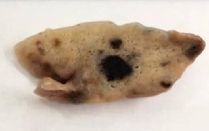


Lung tissue samples from 47 people who died as a result of severe acute respiratory distress syndrome caused by the novel coronavirus were analyzed by Brazilian researchers. The findings can be used to improve treatment of the disease (microscope image of lung infected by SARS-CoV-2, with signs of clotting; credit: Alexandre Fabro/FMRP-USP)
Published on 10/04/2021
By Karina Toledo | Agência FAPESP – COVID-19 can impair the functioning of several organs and has therefore been considered a systemic disease. Even when only a small proportion of patients with respiratory failure are analyzed, it is possible to see that SARS-CoV-2 affects the lungs in different ways.
In a study reported in a paper posted to the preprint platform medRxiv and not yet peer-reviewed, researchers at the University of São Paulo (USP) in Brazil and collaborators analyzed lung tissue samples from 47 people who died as a result of severe acute respiratory distress syndrome (SARDS) caused by the novel coronavirus and identified two distinct patterns of injury.
Five patients (10.6%) displayed what the authors call a “fibrotic phenotype”, characterized by thickening of the alveolar septum (the wall between alveoli, serving as the structural basis for gas exchange in the lungs). In other words, normal tissue in lungs damaged by the virus was replaced by fibrosis (scar tissue), and this hindered breathing. In ten patients (21.2%) with a “thrombotic phenotype”, lung tissue was practically normal, but there were signs of thrombosis (blood clots) in small vessels. A third group comprising 32 patients (68.1%) displayed both phenotypes simultaneously.
The average age of the patients included in the study was 67.8, with similar proportions of men and women. All had pre-existing diseases, mainly high blood pressure (55%) and obesity (36%). At the time of hospital admission, 66% complained of shortness of breath. The main clinical complications observed during their hospital stay were septic shock (62%), acute kidney failure (51%), and acute respiratory distress syndrome (45%).
Lung tissue samples were obtained by minimally invasive biopsy and fixed in formalin and paraffin. They were cut up into sections with a thickness of 3 micrometers (µm, where 1 µm equals a millionth of a meter), stained, and analyzed by microscopy and immunohistochemistry (a technique that entails the use of antibodies against target proteins, such as collagen). RNA from SARS-CoV-2 was identified in all samples using RT-PCR.
“Our starting point was an assessment of lung morphology. Next, we reviewed the patient’s clinical history and radiological exams. After a statistical analysis, we found that the data correlated,” pathologist Alexandre Fabro, last author of the article, told Agência FAPESP. Fabro is a professor at the University of São Paulo’s Ribeirão Preto Medical School (FMRP-USP). The study was supported by FAPESP via three projects (19/01517-3, 19/19591-5, and 20/13370-4).
Respiratory pattern
In the article, the authors report that during the last few days before they died, the fibrotic phenotype patients suffered a gradual decline in oxygenation measured by the ratio of arterial oxygen partial pressure to fractional inspired oxygen (PaO2/FiO2), loss of pulmonary compliance (the lung’s ability to expand and contract during respiration), and augmented production of lung collagen, one of the main components of fibrotic tissue.
In the thrombotic phenotype patients, respiratory patterns improved shortly before they died and compliance was high throughout their stay in hospital. “In some cases, the physician said they came close to being discharged and then died,” Fabro said.
On the other hand, this group was found to have augmented levels of platelets (blood cells involved in clotting) and thrombosis. In addition, they were found on admission to have higher levels of D-dimer, a protein used as a marker of clotting disorder, than the average for the patients analyzed.
“These findings underscore the idea that responses to the virus vary considerably even though the infection is the same, and even among severe cases. This can have clinical implications,” Fabro said. “The results suggest that patients in each group need different treatments. In the article, we show that an analysis of respiratory parameters [PaO2/FiO2] and D-dimer levels at admission, for example, can help medical teams distinguish between these phenotypes.”
The study shows how the process of pulmonary fibrosis that has left many survivors of COVID-19 with long-term symptoms progresses. “One of the most burning scientific questions today is how to treat the disease so that this process doesn’t become permanent,” he said. “There are a few anti-fibrotic drugs, but they haven’t been tested on post-COVID-19 patients.”
The article “COVID-19 bimodal clinical and pathological phenotypes” is at: www.medrxiv.org/content/10.1101/2021.09.03.21262841v1.
Source: https://agencia.fapesp.br/37001