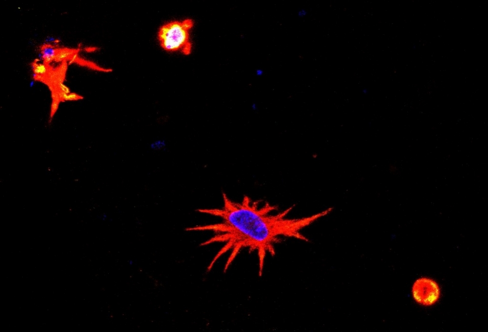


Morphology of a bioprinted astrocyte: cell nucleus stained blue and remainder stained red show a very similar state to that seen in neural tissue (credit: researchers’ archive)
Published on 01/31/2022
By André Julião | Agência FAPESP – A group of researchers affiliated with the Federal University of São Paulo (UNIFESP) in Brazil has developed a protocol for three-dimensional (3D) printing of neural cells. The bioink consisted of natural polymers that enabled astrocytes (a type of brain cell) to survive for at least 14 days in the laboratory after passing through a 3D printer. The procedure resulted in a model more similar to neural tissue than the material obtained by existing protocols, which grow cells in two dimensions.
The study was supported by FAPESP. An article reporting the results is published in the Journal of Visualized Experiments (JoVE).
“In the organism, cells are three-dimensional, but when grown in the laboratory they’re on top of plastic and underneath culture medium [a set of substances that permit cell survival and proliferation]. That’s very different from the natural organization of tissue or organs, where cells are arrayed in three dimensions. The bioink we developed attempts to reproduce the relationship between the cell and the microenvironment and other cells. It’s an intermediate system between 2D culture and experiments with animals,” said Marimélia Porcionatto, a professor at UNIFESP’s Medical School (EPM) and last author of the article.
Astrocytes, the largest and most abundant cells in the central nervous system, play a key role in several neurological processes and in diseases that affect the brain. The procedure standardized by the UNIFESP group can be adapted for the purposes of studying other cell types. The group is currently using it to analyze astrocytes and neurons infected by SARS-CoV-2, the virus that causes COVID-19, as part of another project funded by FAPESP.
“We’re testing different biomaterials for compatibility with neural tissue cells – neurons and neural stem cells, as well as astrocytes. Bioprinting is a recent technique in tissue engineering, and neural tissue cells are particularly sensitive, so this protocol will be useful both for researchers working with astrocytes and other brain cells and for those working with other cell types,” said Bruna Alice Gomes de Melo, first author of the article. The study was conducted during her postdoctoral research at EPM-UNIFESP (more at: agencia.fapesp.br/32523).
The group led by Porcionatto developed the protocol with murine cells but included biocompatible materials that can be adapted for use in studies with human cells. In addition to studies of central nervous system diseases in a format closely resembling that of the brain, the researchers are looking for materials that can be used in future to repair brain areas damaged by traumatic brain injury or stroke, for example (more at: agencia.fapesp.br/29865).
Recipe
The bioink is made from commercially available raw materials such as laminin, a component of the extracellular matrix (molecules located between cells), in this case extracted from cows. The recipe also includes growth factors that enable the cells to survive and thrive in the culture medium.
Another important ingredient is gelatin methacryloyl (GelMA), a material that has proven to be versatile for tissue engineering, drug delivery and 3D printing applications. It is marketed abroad, but the researchers produced it in the laboratory at a far lower cost than imported GelMA. Melo was trained to produce it while she was doing PhD research at the University of Campinas (UNICAMP) in São Paulo state, and during an internship under the Harvard-MIT Program in Health Sciences and Technology (HST) in the United States, with a scholarship from FAPESP.
“In other compositions, many of the cells survived the stress of 3D printing for a time, but astrocyte morphology wasn’t compatible with living tissue. GelMA and laminin were essential,” Melo said.
After the gel-like bioink passes through the printer’s ejector nozzle, it is deposited in layers. A few days later, the astrocytes begin to replicate and behave in a similar manner to that seen in nerve tissue.
The goal now is to increase the complexity of the protocol. As well as astrocytes, the study relating to SARS-CoV-2 used a bioink with neurons and a third material combining both cell types. The researchers plan to include neural stem cells in the mixture in the near future.
“The idea is to get as close as possible to the complexity of neural tissue,” Porcionatto said. “When these protocols are fully validated with mouse cells, we’ll be able to develop others with human cells. They’ll be useful for a variety of studies, such as candidate drug trials, tests to identify genes expressed during brain development, and disease modeling, among others.”
The other co-authors were also researchers at EPM-UNIFESP: Elisa M. Cruz, with a PhD scholarship from FAPESP; Taís N. Ribeiro, with a master’s scholarship; and Mayara Mundim, also with a PhD scholarship.
The article “3D bioprinting of murine cortical astrocytes for engineering neural-like tissue” is at: www.jove.com/t/62691/3d-bioprinting-murine-cortical-astrocytes-for-engineering-neural-like.
Source: https://agencia.fapesp.br/37839