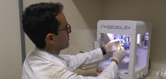
Technique developed at Human Genome and Stem Cell Research Center, funded by FAPESP and hosted by the University of São Paulo, produced hepatic tissue in the laboratory in only 90 days and could become an alternative to organ transplantation in future (photo: Daniel Antônio / Agência FAPESP)
Published on 05/04/2021
By Maria Fernanda Ziegler | Agência FAPESP – Using human blood cells, Brazilian researchers have succeeded in obtaining hepatic organoids (“mini-livers”) that perform all of the liver’s typical functions, such as producing vital proteins, storing vitamins, and secreting bile, among many others. The innovation permits the production of hepatic tissue in the laboratory in only 90 days and may in the future become an alternative to organ transplantation.
The study was conducted at the Human Genome and Stem Cell Research Center (HUG-CELL). Hosted by the University of São Paulo (USP), HUG-CELL is one of the Research, Innovation and Dissemination Centers (RIDCs) funded by FAPESP.
This study combined bioengineering techniques, such as cell reprogramming and the cultivation of pluripotent stem cells, with 3D bioprinting. Thanks to this strategy, the tissue produced by the bioprinter maintained hepatic functions for longer than reported by other groups in previous studies.
“More stages have yet to be achieved until we obtain a complete organ, but we’re on the right track to highly promising results. In the very near future, instead of waiting for an organ transplant, it may be possible to take cells from the patient and reprogram them to make a new liver in the laboratory. Another important advantage is zero probability of rejection, given that the cells come from the patient,” said Mayana Zatz, director of HUG-CELL and last author of the article published in Biofabrication.
The innovative part of the study resided in how the cells were included in the bioink used to produce tissue in the 3D printer. “Instead of printing individualized cells, we developed a method of grouping them before printing. These ‘clumps’ of cells, or spheroids, are what constitute the tissue and maintain its functionality much longer,” said Ernesto Goulart, a postdoctoral fellow in USP’s Institute of Biosciences and first author of the article.
The researchers thereby avoided a problem faced by most human tissue bioprinting techniques, namely, the gradual loss of contact among cells and hence loss of tissue functionality.
Spheroid formation in this study already occurred in the differentiation process, when pluripotent cells were transformed into hepatic tissue cells (hepatocytes, vascular cells, and mesenchymal cells). “We started the differentiation process with the cells already grouped together. They were cultured in agitation, and groups formed spontaneously,” Goulart told Agência FAPESP.
A liver in 90 days
According to the researchers, the complete process from collection of the patient’s blood to functional tissue production takes approximately 90 days and can be divided into three stages: differentiation, printing, and maturation.
Initially, the blood cells are reprogrammed to regress to a stage of pluripotency characteristic of stem cells, becoming induced pluripotent stem cells (iPSCs). Japanese scientist Shinya Yamanaka was awarded the 2012 Nobel Prize for Medicine for developing this technique.
The next stage consists of inducing differentiation into liver cells. The spheroids are then mixed with bioink, a hydrogel-like fluid, and printed out. The resulting structures mature in culture for 18 days.
“The printing process entails the deposition of spheroids along three axes, which is necessary for the material to gain volume and give the tissue proper support,” Goulart said. “The gel-like bioink is crosslinked to make the structures more rigid so that they can be manipulated and even sutured.”
Most of the available methods for printing live tissue use immersion and cell dispersion in a hydrogel to recapitulate the microenvironment and ensure tissue functionality. However, experiments have shown that loss of cell contact and functionality tends to occur when dispersion is performed cell by cell.
“It’s a somewhat traumatic process for the cells, which need time to get used to the environment and acquire functionality,” Goulart said. “At this stage, they aren’t tissue yet because they’re dispersed, but as shown by our study, they already have the capacity to clear the blood of toxins and to produce and secrete albumin [a protein produced only by the liver], for example.”
In this study, researchers developed mini-livers using blood cells from three volunteers as raw material and compared markers relating to functionality, such as the maintenance of cell contact and protein production and release. “Our spheroids worked much better than those obtained from single-cell dispersion. As expected, during maturation, the markers of hepatic function were not reduced,” Goulart said.
Although the study was limited to producing miniature livers, the technique can be used in the future to produce complete organs suitable for transplantation, according to Goulart. “We did it on a small scale, but with investment and interest, it can easily be scaled up,” he said.
The article “3D bioprinting of liver spheroids derived from human induced pluripotent stem cells sustain liver function and viability in vitro” (doi.org/10.1088/1758-5090/ab4a30) by Ernesto Goulart, Luiz Carlos de Caires-Junior, Kayque Alves Telles-Silva, Bruno Henrique Silva Araujo, Silvana Aparecida Rocco, Mauricio Sforca, Irene Layane de Sousa, Gerson Shigeru Kobayashi, Camila Manso Musso, Amanda Faria Assoni, Danyllo Oliveira, Elia Caldini, Silvano Raia, Peter I. Lelkes and Mayana Zatz can be retrieved from https://iopscience.iop.org/article/10.1088/1758-5090/ab4a30.
Source: https://agencia.fapesp.br/32217
