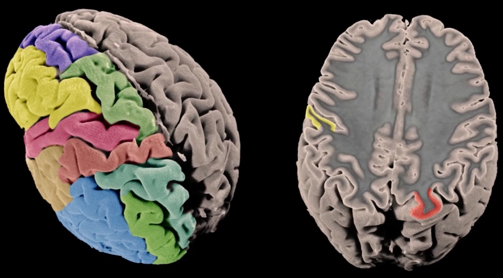


Based on quantitative MRI, researchers measured cortical volume in 51,665 participants and correlated the data with genetic markers (image: Tyler Ard, James Stanis & Arthur Toga / Stevens Neuroimaging and Informatics Institute, Keck School of Medicine of the University of Southern California)
Published on 03/23/2021
By Karina Toledo | Agência FAPESP – Mapping small variations in the human genome that may influence the architecture of the cerebral cortex and correlate with a predisposition to diseases such as schizophrenia, epilepsy, autism, bipolar disorder, anorexia, depression and dementia, among others, was the purpose of a study involving more than 360 scientists affiliated with 184 institutions around the world. The findings are published in the journal Science.
In an analysis of quantitative brain magnetic resonance imaging (MRI) data from 51,665 individuals in various countries, the scientists identified 306 genetic variants thought to influence the structure of key brain regions. The investigation was conducted under the aegis of ENIGMA (Enhancing Genetic Neuroimaging through Meta-Analysis), an international consortium dedicated to studying several neurological and psychiatric diseases. Its members include the Brazilian Institute of Neuroscience and Neurotechnology (BRAINN) at the University of Campinas (UNICAMP) in the state of São Paulo. BRAINN is one of the Research, Innovation and Dissemination Centers (RIDCs) supported by FAPESP.
“It’s the most comprehensive neuroimaging study of the cerebral cortex ever conducted, mapping the genetic architecture of the human brain for the first time,” Fernando Cendes, a professor at UNICAMP and coordinator of BRAINN, told Agência FAPESP.
According to Cendes, the main advantage of such a large amount of information is that it can be used to detect even very discreet alterations in brain structure that would not otherwise be perceptible. “The findings give us a better understanding of the functioning and structure of the brain, both healthy and diseased,” he said.
Two kinds of analysis
The analyses described in the Science article were based on two large datasets. The first comprised quantitative brain MRI scans, which the researchers used to calculate cortical volume. Approximately 25,000 participants were healthy and served as controls. The remaining were patients with conditions such as insomnia, depression, attention-deficit/hyperactivity disorder (ADHD), epilepsy, and Parkinson’s disease.
“The cerebral cortex is the outermost layer of the brain, made up of folded gray matter full of grooves [sulci] and ridges [gyri],” Cendes said. “It’s rich in neurons and responsible for high-order cognitive functions such as language, emotion, memory and information processing.”
In the study, the cerebral cortex was divided into 34 relatively homogeneous regions. Two parameters were measured for each region: cortical thickness (the distance between the white matter surface and the pial surface below the dura mater, the membrane that surrounds the brain) and cortical area.
“All groups used a special technique to acquire high-resolution three-dimensional images,” Cendes said. “Measurements were made automatically with the aid of computational algorithms. This is important because it eliminates examiner bias.”
The other dataset comprised whole-genome sequences of the participants and tissue samples deposited with brain banks, enabling the researchers to analyze genetic markers such as single nucleotide polymorphisms (SNPs), which are variations in DNA sequences that affect only one base (adenine, cytosine, guanine or thymine) and can be used to compare different individuals’ genomes.
The next step consisted of correlating the cortical measurements with the genetic variations detected and then comparing the patterns observed in healthy subjects with those found in individuals with different symptoms and diseases.
Each research group analyzed the data for its local patients and controls. A meta-analysis was coordinated by Katrina Grasby and other members of the Psychiatric Genetics Research Group at QIMR Berghofer Medical Research Institute in Australia, in collaboration with researchers at the University of Southern California and the University of North Carolina at Chapel Hill in the United States. BRAINN contributed data for 150 Brazilians.
Last, they produced a kind of map identifying the brain regions with augmented or diminished volume in a person with epilepsy, for example, and the genes correlated with these alterations.
“The results enhance our understanding of the genes associated with the development of each of the 34 regions of the cortex we studied. They show that changes in the architecture of the cerebral cortex as well as genetic variants can predispose individuals to certain diseases,” Cendes said.
According to Grasby, the study shows that the genetic variations associated with reductions in cortical volume also contribute to a higher risk of ADHD, depression and insomnia. “This gives us a starting point for further exploration of the genetic links between brain structure and disease,” she said.
Correlations were also found between certain genes or cortical areas and cognitive performance or educational attainment, Cendes added.
“These correlations are by no means 100%. Several other genes are associated with a predisposition to each of the diseases or cognitive functions investigated and haven’t been identified,” he said. “Nevertheless, based on this first study, it’s possible to move on to a broader analysis using artificial intelligence to identify potential biomarkers for complex diseases that affect the brain.”
The group’s final objective is understanding how genes modulate brain structure and identifying clusters of genetic alterations that can predict whether a person is more likely to develop a certain disease.
“This would be a step toward personalized medicine,” Cendes said. “If we know how brains differ from one another, we can design the right treatment strategy for each person and even advise patients on specific preventive measures.”
The article “The genetic architecture of the human cerebral cortex” can be retrieved from science.sciencemag.org/content/367/6484/eaay6690.
Source: https://agencia.fapesp.br/33403